|
skin structure professionals
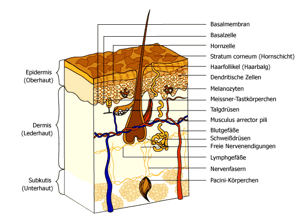
Epidermis
This layer forms the external surface of the skin. It is mainly composed of keratinocytes, which are stacked on top of each other, forming 4 sub-layers (basal layer, spinous layer, granular layer and surface layer). The keratinocytes develop at the bottom and rise to the top, where they lose their nucleus and are finally shed from the surface. This migration process takes at least 28 days. The epidermis is constantly renewing itself, the live cells changing into dead, hard, flattened cells. Other cellular components of the epidermis are melanocytes and Langerhans cells
- Melanocytes
These cells produce melanin for skin pigmentation which partially provides UV protection. They are located at the bottom of the epidermis
- Dendritic (Langerhans) cells
These are dendritic antigen-presenting cells involved in the epidermal immune system. They engulf foreign material that invades the epidermis and migrate out of the skin to stimulate an immune response.
Dermis
The dermis is separated from the epidermis by the basement membrane and consists mostly of connective tissue. It is divided into 2 layers, the papillary dermis and the reticular dermis. It is responsible for the skin's pliability and mechanical resistance and is also involved in the regulation of the body temperature. The dermis supplies the avascular epidermis with nutrients by means of its vascular network. It contains sense organs for touch, pressure, pain and temperature (Meissner´s corpuscles, Pacinian corpuscles, free nerve endings), as well as blood vessels, nerve fibres, sebaceous and sweat glands and hair follicles.
- Meissner's corpuscle
These touch receptors are expecially effective in detecting light touch and soft, fleeting movements.
- Pacinian corpuscles
Pacinian corpuscles function as receptors for deep pressure and vibration.
- Free Nerve Endings
Free nerve endings are sensitive to pain, temperature changes and itchiness.
- Nerve Fibers
Nerve fibres forward information.
- Sebaceous Glands
Sebaceous or oil glands are small, sacculated organs that secrete sebum. This oily substance is a natural moisturiser which conditions the hair and skin. Sebaceos glands are found all over the body, but they are more numerous in the scalp area and around the forehead, chin, cheeks and nose.
- Sweat Glands
These are sweat-producing structures consisting of a single tube, a coiled body and a superficial duct. They are involved in thermoregulation, as they cool the skin by sweating.
- Hair Follicles
Hair follicles are downward growths into the dermis of epidermal tissue and produce hair. They are found all over the body except on the palms and soles as well as on the lips.
- Arrector pili muscle
This small muscle is attached to the base of the follicle. When it is stimulated by cold or fright, it pulls the hair follicle up, causing it to stand upright.
Subcutaneous layer
This layer consists mainly of adipose tissue, surrounded by loose connective tissue, and serves to attach the dermis to its underlying tissues. Not all authors see this layer as a part of the skin, but it definitely has a strong impact on the way the skin looks.
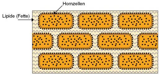
The stratum corneum (horny layer)
This thin, most superficial layer of the skin forms the interface with the external environment. Knowledge of its structure, composition and function provides insights into the basis of dry and scaly skin disorders, possibly leading to increasingly functional topical therapies.
The stratum corneum is composed of terminally differentiated keratinocytes called corneocytes, surrounded by a matrix of specialised lipids. It can be likened to a brick wall, composed of anucleated cells (the "bricks") and intercellular lamellar membranes (the "mortar").
The corneocytes are filled with structural proteins (keratin filaments) and osmotically active small molecules, they do not contain any organelles. The corneocyte cytosol is encased by a chemically resistant protein shell, the cornified cell envelope.
The major lipid species of the stratum corneum are ceramides, essential and non-essential fatty acids and cholesterol. In contrast to any other known biological membrane, phospholipids are absent.
The primary function of the stratum corneum is to retard evaporative water loss from the interior. Besides, its barrier properties protect from ultraviolet radiation, oxidants, microorganisms and toxic agents.
|