|
skin structure
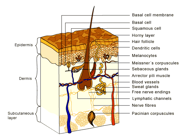
The skin consists of three main layers epidermis, dermis and subcutis. Each layer has its specific structure and functions.
Epidermis
This layer is seen on the surface of the skin. It is made up of cells called keratinocytes, which are stacked on top of each other, forming different sub-layers. The keratinocytes develop at the bottom and rise to the top, where they are shed from the surface as dead cells. So this layer is constantly renewing itself, the live cells changing into dead, hard, flattened cells. Melanocytes and Langerhans cells are other important cells found in the epidermis which have special functions
- Melanocytes
These cells produce a dark pigment called melanin which contributes to skin color and provides UV protection. They are located at the bottom of the epidermis.
- Dendritic (Langerhans) cells
These cells are involved in the epidermal immune system. They engulf foreign material that invades the epidermis and migrate out of the skin to stimulate an immune response.
- Basal cells
Small cells found at the bottom of the epidermis. Earlier it was believed that basal cell carcinoma is derived from these cells. As of this writing basal cell carcinoma is thought to arise from non-differentiated cells from the basal cell layer.
Dermis
The dermis consists mostly of connective tissue and is much thicker than the epidermis. It is responsible for the skin's pliability and mechanical resistance and is also involved in the regulation of the body temperature. The dermis supplies the avascular epidermis with nutrients by means of its vascular network. It contains sense organs for touch, pressure, pain and temperature (Meissner´s corpuscles, Pacinian corpuscles, free nerve endings), as well as blood vessels, nerve fibres, sebaceous and sweat glands and hair follicles.
- Blood Vessels
These are tiny pipes through which blood circulates. The blood vessels supply the skin with fresh blood, which contains nutrients and oxygen, and carry away waste products.
- Meissner's corpuscle
These touch receptors are expecially effective in detecting light touch and soft, fleeting movements.
- Pacinian corpuscles
Pacinian corpuscles function as receptors for deep pressure and vibration.
- Free Nerve Endings
Free nerve endings are sensitive to pain, temperature changes and itchiness.
- Nerve Fibers
Nerve fibres forward information.
- Sebaceous Glands
Sebaceous or oil glands are small, sacculated organs that secrete sebum. This oily substance is a natural moisturiser which conditions the hair and skin. Sebaceos glands are found all over the body, but they are more numerous in the scalp area and around the forehead, chin, cheeks and nose.
- Sweat Glands
These are sweat-producing structures consisting of a single tube, a coiled body and a superficial duct. They are involved in thermoregulation, as they cool the skin by sweating.
- Hair Follicles
Hair follicles are downward growths into the dermis of epidermal tissue and produce hair. They are found all over the body except on the palms of the hands and soles of the feet as well as on the lips. When the body gets cold, the hair stands upright with the help of the arrector pili muscle, closing up the skin's pores and keeping the warmth in.
- Arrector pili muscle
This small muscle is attached to the base of the follicle. When it is stimulated by cold or fright, it pulls the hair follicle up, causing it to stand upright.
Subcutaneous layer
The subcutaneous layer below the dermis consists of loose connective tissue and much fat. It acts as a protective cushion and helps to insulate the body by monitoring heat gain and heat loss. Not all authors consider this layer a part of the skin, but it definitely has a strong impact on the way the skin looks.
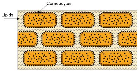
The horny layer
This thin, most superficial layer of the skin forms the interface with the external environment and has special protective functions
It is made of dead, flat skin cells, surrounded by specialised fatty substances (lipids). It can be likened to a brick wall, composed of cells (the "bricks") and lipids between the cells (the "mortar").
The primary function of the horny layer is to hinder water loss through evaporation from the interior. Besides, it protects from ultraviolet radiation, mechanical damage, foreign chemicals and germs.
|